
Make an appointment
When it comes to your health, you deserve quality service. You can rely on our team for your medical examinations. To make an appointment, contact our team by phone, fax or click the “Make an appointment” link.
819 820-2827
Fax: 819 563-3542
Ultrasound and Doppler
Echography, or ultrasonography, is a medical imaging method that uses sound waves to produce detailed images of the body’s interior. Ultrasound waves bounce off organs to create a multi-dimensional image.
Description
Painless, non-invasive and radiation-free, it is essential in many medical contexts to diagnose and monitor various conditions.
Doppler tests are complementary tools that can also be used to analyze blood circulation.
Why have an ultrasound?
Ultrasound is commonly used to:
- Observe internal organs such as the liver, kidneys, gallbladder and thyroid.
- Monitor pregnancies and evaluate fetal development.
- Detect soft tissue abnormalities such as cysts or tumours.
- Assess blood circulation using advanced techniques, such as Doppler.
- Guide medical interventions such as biopsies or infiltrations.
Types of Exams Offered
- Pelvic and Abdominal Ultrasound
- Cardiac Ultrasound
- Breast Ultrasound
- Testicular Ultrasound
- Musculoskeletal Ultrasound
- Neck/Thyroid Ultrasound
- Upper and Lower Extremity Ultrasound
- Endovaginal Ultrasound
- Venous or Carotid Doppler
- Renal and Abdominal Doppler
Preparation for the test
Preparation for ultrasound procedures may vary depending on the type of ultrasound.
Abdomen or bladder: Fast 6 to 8 hours before the examination.
Pelvis: Drink 1 to 1.5 litres of water at least one hour before the procedure. Do not urinate before the test; a full bladder is necessary.
Other ultrasounds: No specific preparation, unless otherwise indicated.
Our technologists will give you all the necessary instructions before your appointment.
At the time of your appointment, please make sure you have:
- your valid health insurance card
- the original medical prescription signed by the requesting professional
Ultrasound procedure
Preparation: Depending on the type of ultrasound (abdominal, pelvic, etc.), specific instructions (fasting, full bladder) may be necessary.
Patient position: You will be lying down or placed in a suitable position to allow optimal access to the area being examined.
Gel application: A conductive gel is applied to the skin to facilitate sound wave transmission.
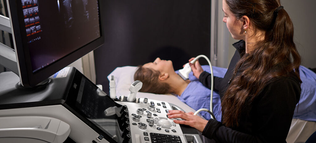
Use of the probe: The technician or radiologist moves the probe (transducer) over the area being examined to capture images in real time.
Duration: Approximately 15 to 30 minutes, depending on the complexity of the procedure.
Doppler procedure
Doppler generally follows the same steps as an ultrasound, with a few specific differences:
Duration: May be slightly longer than a standard ultrasound, depending on the vascular segments studied (20 to 40 minutes).
Preparation and position: Identical to ultrasound, but the patient’s position may need to be altered slightly to optimize access to the targeted vessels.
Use of a Doppler probe: Similar to the ultrasound probe, but this captures additional information on blood flow using the Doppler effect (analysis of frequency changes in reflected sound waves).
Visualization of blood flow: In real time, colours or graphs indicate the speed and direction of blood flow in the vessels.
Precautions and contraindications
Ultrasound is generally risk-free, but a few precautions should be noted:
Open wounds: If the area being examined is injured, this may affect the examination or require adjustments.
Pregnancy and breastfeeding: Totally safe, including Doppler ultrasounds.
Pacemakers or medical implants: Let us know if you have any implanted devices that could influence certain examinations.
Fees
Our ultrasound and Doppler examination services are covered by the Régie de l’assurance maladie du Québec (RAMQ).
To view the fees for all the procedures we perform, please consult our fee schedule. You can also check whether procedure fees are covered by the RAMQ.
FAQ
Would you like to know more about ultrasound and Doppler exams? Consult our frequently asked questions to find out the answers to the most frequently asked questions about these non-invasive examinations.
How long does an ultrasound take?
An ultrasound can take between 15 and 30 minutes, depending on the area being examined and the complexity of the exam.
Why is it necessary to have a full bladder for an ultrasound?
During a pelvic or abdominal ultrasound, a full bladder helps push the intestines aside and provides a clear view of the pelvic organs and the abdominal wall. In addition, the fluid in the bladder transmits ultrasound waves very well, which improves image clarity.
However, if the exam requires an endovaginal ultrasound, you will be asked to completely empty your bladder beforehand.
What can be detected with an ultrasound?
Ultrasound allows for the visualization of internal organs and soft tissues and the detection of abnormalities such as cysts, masses, stones, or inflammation, without the use of X-rays. Among other things, it can be used to examine the abdominal and pelvic organs, monitor fetal development during pregnancy, distinguish between tumors and cysts, assess blood flow (Doppler ultrasound), examine the thyroid and lymph nodes, and detect muscle tears and joint inflammation.
What can’t be seen on an ultrasound?
On an ultrasound, bone structures and air-filled organs, such as the lungs or intestines, are not visible because ultrasound waves cannot pass through them. Certain microscopic fetal abnormalities may also go undetected by ultrasound.
What is the difference between an X-ray and an ultrasound?
The main difference lies in the technology used: X-rays use ionizing radiation, while ultrasounds use high-frequency sound waves (ultrasonics). Each of these tests plays a specific role depending on what you’re trying to observe.
X-rays are ideal for viewing bones and lungs, whereas ultrasound allows for the observation of organs and tissues and for monitoring fetal development.
What is a Doppler exam?
A Doppler exam is a type of ultrasound that uses sound waves to observe blood flow in arteries and veins. It assesses the volume, speed, and direction of blood flow to detect any blockages or narrowing of the blood vessels.
Why have a Doppler ultrasound?
A Doppler ultrasound can be used to diagnose many vascular conditions, such as phlebitis (thrombosis) and arterial diseases, and to screen for cholesterol plaques in the neck arteries to help prevent the risk of stroke.
How long does a Doppler last?
A Doppler scan can be slightly longer than a standard ultrasound, between 20 and 40 minutes depending on the vascular segments studied.
What is the difference between a Doppler and an ultrasound?
Ultrasound allows us to visualize the structure of organs and tissues, while Doppler specifically evaluates blood circulation in the vessels. Both examinations use ultrasound, but fulfill complementary objectives. The Doppler is an additional module that can be added to a standard ultrasound. We first use the ultrasound image to see the vessel, then the Doppler function allows us to analyze the blood circulating there.
Any questions?
Contact us